The heart is a vital organ and a central topic in GCSE Biology. It is essential for understanding the circulatory system and how blood transports oxygen and nutrients throughout the body. Its role in maintaining life makes it a crucial focus for exams, where students are often tested on their ability to label and explain the heart’s structure and functions. So what we learn about the heart diagram GCSE biology?
Understanding the heart’s anatomy and how it works is not just important for scoring well on exams but also provides important knowledge for further studies in biology and healthcare. Here we offer a detailed explanation of the heart’s structure. You can learn about the pathway of blood flow, and its role in the human body. Let’s dive into the intricate workings of the human heart and master this essential GCSE Biology topic!
What Is A Structure of the Heart: A Detailed Overview
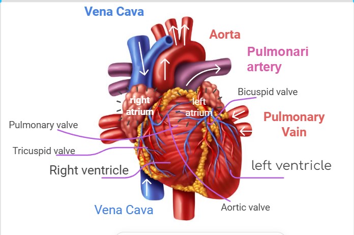
The human heart is a muscular organ that functions as a pump to circulate blood throughout the body. It devides into several distinct anatomical features, each playing a vital role in the circulatory system. Understanding these components is essential for mastering the heart diagram in GCSE Biology.
Chambers of the Heart
The heart has four chambers that work together to pump blood:
Right Atrium: Receives deoxygenated blood from the body through the vena cava.
Right Ventricle: Pumps deoxygenated blood to the lungs via the pulmonary artery for oxygenation.
Left Atrium: Receives oxygenated blood from the lungs through the pulmonary vein.
Left Ventricle: Pumps oxygenated blood to the rest of the body via the aorta.
The left ventricle has the thickest muscular wall because it must generate enough force to pump blood throughout the entire body.
Valves of the Heart
Valves ensure one-way blood flow and prevent backflow.
Tricuspid Valve: Between the right atrium and right ventricle.
Bicuspid (Mitral) Valve: Between the left atrium and left ventricle.
Pulmonary Valve: Between the right ventricle and pulmonary artery.
Aortic Valve: Between the left ventricle and aorta.
These valves open and close with each heartbeat, maintaining efficient blood circulation.
Major Blood Vessels
Aorta: Carries oxygenated blood from the left ventricle to the body.
Vena Cava: Brings deoxygenated blood from the body to the right atrium.
Pulmonary Artery: Transports deoxygenated blood from the right ventricle to the lungs.
Pulmonary Vein: Brings oxygenated blood from the lungs to the left atrium.
These vessels connect the heart to the lungs and the rest of the body, completing the circulatory loop.
Muscular Walls
The ventricles have thicker muscular walls compared to the atria because they pump blood over longer distances.
The left ventricle is the most muscular, as it pumps blood to the entire body, while the right ventricle only pumps blood to the lungs.
The Heart Diagram GCSE – The Blood Flow Through the Heart
Now let’s learn about how the blood flows through the heart. The heart’s primary function is to pump blood through the body. It ensures that oxygen and nutrients are delivered to cells while removing carbon dioxide and waste products. Understanding the flow of blood through the heart is essential for you to master GCSE Biology.
Deoxygenated Blood Enters the Heart
Blood returning from the body is low in oxygen and high in carbon dioxide. Vena Cava: Deoxygenated blood enters the heart through two large veins: Superior Vena Cava: Drains blood from the upper body. Inferior Vena Cava: Drains blood from the lower body. Blood flows into the right atrium, the first chamber it encounters in the heart.
Right Atrium to Right Ventricle
- From the right atrium, blood passes through the tricuspid valve into the right ventricle.
- The tricuspid valve prevents blood from flowing back into the atrium when the ventricle contracts.
Blood Travels to the Lungs
- The right ventricle pumps deoxygenated blood into the pulmonary artery through the pulmonary valve.
- The pulmonary artery carries the blood to the lungs, where it undergoes gas exchange:
- The Blood releases Carbon dioxide.
- Blood absorbs An Oxygen.
Oxygenated Blood Returns to the Heart
Oxygen-rich blood returns to the heart via the pulmonary vein. It flows into the left atrium, marking the start of its journey through the oxygenated part of the circulatory system.
Left Atrium to Left Ventricle
From the left atrium, blood passes through the bicuspid (mitral) valve into the left ventricle. The bicuspid valve ensures that blood flows in one direction, preventing backflow into the left atrium.
Blood is Pumped to the Body
The left ventricle pumps oxygenated blood into the aorta through the aortic valve. The aorta distributes the oxygen-rich blood to the rest of the body, supplying tissues and organs with the oxygen and nutrients they need for survival.
Blood Flow Pathway Summary
Chambers of the Heart
| Chamber | Details | Function |
| Right Atrium | Receives deoxygenated blood from the body via the vena cava. | Passes blood to the right ventricle. |
| Right Ventricle | Pumps deoxygenated blood to the lungs through the pulmonary artery. | Sends blood to the lungs for oxygenation. |
| Left Atrium | Receives oxygenated blood from the lungs via the pulmonary veins. | Passes blood to the left ventricle. |
| Left Ventricle | Pumps oxygenated blood to the body through the aorta. | Supplies oxygenated blood to all body tissues. |
Here’s a simplified step-by-step sequence of blood flow:
Deoxygenated Blood:
Vena cava → Right atrium → Tricuspid valve → Right ventricle → Pulmonary artery → Lungs.
Oxygenated Blood: Pulmonary vein → Left atrium → Bicuspid valve → Left ventricle → Aorta → Body.
The Double Circulation System In The Heart Diagram GCSE
The human circulatory system efficiently delivers oxygen and nutrients precisely where they’re need to be and effectively removes waste products like carbon dioxide. The double circulation system makes this possible by allowing blood to pass through the heart twice during each complete circuit of the body. This system consists of two distinct pathways: pulmonary circulation and systemic circulation.
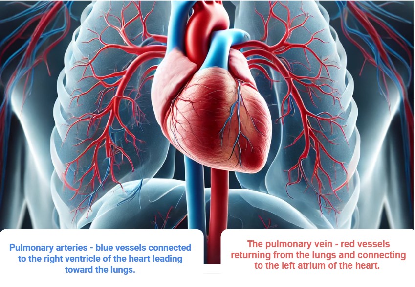
Pulmonary Circulation: The Journey to the Lungs
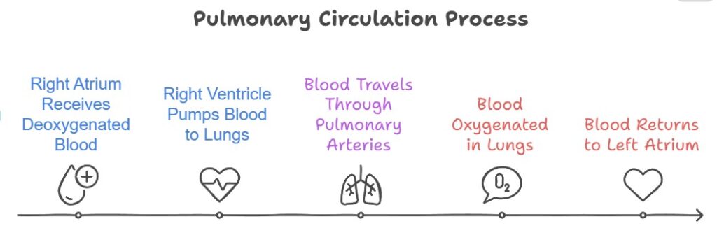
Pulmonary circulation focuses on the exchange of gases between the heart and lungs. Here’s how it works:
Deoxygenated blood from the body enters the right side of the heart (via the vena cava) and is pumped into the pulmonary artery. This blood travels to the lungs, where it picks up oxygen and releases carbon dioxide during gas exchange. The now oxygenated blood returns to the heart through the pulmonary veins, ready for distribution to the rest of the body.
Pulmonary circulation ensures that the blood is recharged with oxygen, which is vital for keeping cells functioning and alive.
Systemic Circulation: Delivering Oxygen and Nutrients
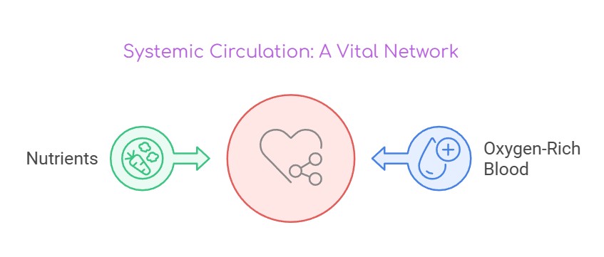
The heart pumps oxygenated blood into the systemic circulation, delivering oxygen and nutrients to the entire body. Here’s how it works:
The left side of the heart receives oxygen-rich blood and pumps it into the aorta, the body’s largest artery. This blood flows through a vast network of arteries, reaching tissues and organs to deliver oxygen and nutrients. After the oxygen is used, veins carry the now deoxygenated blood back to the heart, completing the circuit.
Systemic circulation supports all the cells in your body, enabling them to perform vital functions like energy production, repair, and growth.
Why Is Double Circulation Efficient and Necessary?
- Efficient Oxygen Delivery: By separating oxygenated and deoxygenated blood, double circulation ensures that tissues receive blood rich in oxygen and nutrients, improving overall efficiency.
- High Blood Pressure for the Body: Systemic circulation operates under higher pressure, allowing oxygenated blood to reach distant parts of the body quickly and effectively.
- Low Pressure for the Lungs: Pulmonary circulation operates at lower pressure to protect the delicate tissues in the lungs during gas exchange.
- Maintaining Nutrient Supply: Double circulation ensures a constant supply of oxygen and nutrients to sustain cellular activities, enabling growth, repair, and energy production.
- Effective Waste Removal: Deoxygenated blood efficiently carries carbon dioxide and other waste products to the lungs and kidneys for elimination.
The Heart Diagram GCSE: Functions of the Heart – The Engine of Life
The heart plays a central role in keeping us alive and healthy by acting as a highly efficient pump. It ensures that oxygen, nutrients, and other vital substances reach every cell in the body while removing waste products. Let’s explore the key functions of the heart in detail.
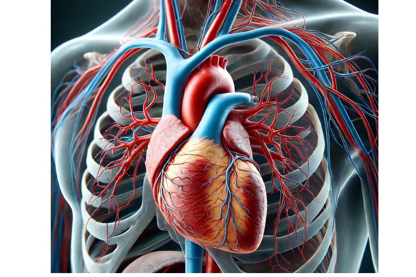
The Heart Structure and Function
| Component | Details | Function |
| Valves | Tricuspid, Pulmonary, Mitral, and Aortic valves prevent backflow of blood. | Ensure unidirectional blood flow. |
| Septum | Muscular wall dividing the left and right sides of the heart. | Prevents mixing of oxygenated and deoxygenated blood. |
| Coronary Arteries | Supply oxygen-rich blood to the heart muscle. | Keep the heart functioning efficiently. |
| Major Blood Vessels | Include the Vena Cava, Pulmonary Artery, Pulmonary Veins, and Aorta. | Transport blood to and from the heart. |
Pumping Oxygenated Blood to Tissues
The heart is responsible for delivering oxygen-rich blood to the body through the systemic circulation system.
After oxygenation in the lungs, blood enters the left atrium and is pumped into the left ventricle. The left ventricle contracts powerfully, sending oxygenated blood into the aorta, the largest artery in the body. From the aorta, blood travels to tissues and organs, supplying the oxygen needed for energy production and cellular functions.
Without this function, tissues would quickly become deprived of oxygen. It will lead to cell death and organ failure.
Removing Carbon Dioxide and Waste Products
The heart also helps rid the body of carbon dioxide and other waste products via pulmonary circulation.
- Deoxygenated blood, carrying carbon dioxide and metabolic waste, is pumped from the right side of the heart to the lungs through the pulmonary artery.
- In the lungs, carbon dioxide is exchanged for oxygen and exhaled out of the body.
- The cleaned, oxygen-rich blood returns to the heart to begin the systemic circuit again.
This cycle ensures that waste products are efficiently eliminated, maintaining a healthy internal environment.
Maintaining Blood Pressure
The heart generates the force needed to maintain blood pressure, which is crucial for ensuring blood reaches all parts of the body.
- The left ventricle, with its thick muscular walls, creates the high pressure needed to push blood through the systemic circuit.
- Blood pressure allows nutrients, oxygen, and hormones to diffuse effectively into tissues and cells.
If blood pressure falls too low, organs may not receive enough blood flow, while excessively high blood pressure can damage blood vessels and organs.
Enabling the Double Circulation System
The heart’s structure and function are integral to the double circulation system, which separates oxygenated and deoxygenated blood:
The right side of the heart handles pulmonary circulation, sending deoxygenated blood to the lungs.
The left side of the heart manages systemic circulation, pumping oxygen-rich blood to the body.
This separation ensures efficiency, allowing the heart to deliver oxygen and nutrients where they’re needed most while removing carbon dioxide and waste.
Exam Relevance: The Heart Diagram GCSE Biology
The heart diagram is a key topic in GCSE Biology exams, and students are often tested on their understanding of its structure and function. Exam questions can take various forms, and mastering this topic is essential for achieving high marks. Hear are some exam tips for the heart diagram GCSE, which will help you during preparation.
How the Heart Diagram GCSE Is Tested?
Labeling Tasks
Students may be asked to label parts of the heart on a diagram. This typically involves identifying key structures, such as: Chambers (e.g., left atrium, right ventricle). Valves (e.g., tricuspid, bicuspid). Blood vessels (e.g., aorta, pulmonary vein).
Tip: Practise labelling diagrams regularly, and focus on spelling accuracy and correct placement of terms.
Descriptive Questions
These questions require explaining processes such as blood flow, the role of valves, or how oxygenated and deoxygenated blood is kept separate. For example: Describe how blood flows through the heart and lungs; Explain the function of the tricuspid valve.
Tip: Use clear and concise language. Break answers into logical steps, and include specific terms like “pulmonary artery” or “oxygenated blood.”
Multiple-Choice Questions
These questions test quick recall and understanding of concepts, such as:
The pathway of blood flow through the heart.
The function of valves or major blood vessels.
Characteristics of the double circulation system.
Tip: Read all options carefully before selecting an answer, and eliminate choices you know are incorrect to improve your chances.
Tips for Answering Questions Effectively For Heart Diagram GCSE Biology
Understand the Diagram: Memorise the heart diagram thoroughly. Practise drawing and labelling it from memory to ensure you’re exam-ready.
Master Key Terms: Be familiar with terms like “atria,” “ventricles,” “pulmonary artery,” and “aorta.” Using precise terminology can earn you marks.
Learn Blood Flow Pathways: Know the step-by-step flow of blood through the heart, lungs, and body. This is frequently asked in descriptive questions.
Revise with Past Papers: Practice exam-style questions to familiarise yourself with the format and improve your confidence.
Explain Clearly: When answering descriptive questions, structure your response logically. Use bullet points if necessary to outline steps clearly.
Use Diagrams: If allowed, draw a quick diagram in your answer to support your explanation.
To properly prepare for the heart diagram GCSE and other biology related topics you can use GCSE Biology Past Papers.
Learn About The Heart Diagram GCSE Biology through listening:
Sometimes, after the good read some students prefer effective way of watch and learn. So if you prefer learning about the heart diagram GCSE through listening, you can watch this video below:
Conclusion For The Heart Diagram GCSE Biology
Mastering the heart diagram is essential for GCSE Biology, as it provides the foundation for understanding the circulatory system and its vital role in sustaining life. It important for you to know how to label the heart accurately. Also, explain blood flow pathways, and describe its functions. This prepares you not only for exams but also enhances your overall understanding of human biology.
To succeed, regular practice is key. Repeatedly revising the heart’s structure, practicing exam-style questions is essential. Also, drawing the diagram from memory can significantly boost confidence and accuracy. Additionally, seeking support from online GCSE Biology tutors can provide personalized guidance, helping you clarify complex concepts, refine their answers, and stay on track with their revision.
With dedication and the right resources, you can excel in heart diagram GCSE biology and achieve excellent results in their GCSE exams. We hope our blog was useful for you. Good Luck!
FAQ’s About The Heart Diagram GCSE
What Is the Structure of the Heart in GCSE?
The heart consists of four chambers: the right atrium, left atrium, right ventricle, and left ventricle. It has four valves: tricuspid, bicuspid (mitral), pulmonary, and aortic valves, which ensure one-way blood flow. Major blood vessels include the aorta, vena cava, pulmonary artery, and pulmonary vein. Each component works together to pump blood throughout the body efficiently.
. What Are the 7 Steps of Blood Flow Through the Heart?
The seven steps of blood flow through the heart are:
- The left ventricle pumps oxygenated blood through the aortic valve into the aorta, delivering it to the body.
- Deoxygenated blood enters the right atrium via the vena cava.
- Blood flows through the tricuspid valve into the right ventricle.
- The right ventricle pumps blood through the pulmonary valve into the pulmonary artery.
- Blood travels to the lungs, where it picks up oxygen and releases carbon dioxide.
- Oxygenated blood returns to the left atrium via the pulmonary vein.
- Blood passes through the bicuspid (mitral) valve into the left ventricle.
Why Does the Heart Beat Twice in GCSE?
The heart beats twice per cycle because of its double circulation system, which separates pulmonary and systemic circulation. The first beat pumps deoxygenated blood to the lungs for oxygenation, while the second beat pumps oxygenated blood to the rest of the body. This system ensures efficient oxygen delivery and waste removal.
How Does the Heart Work in IGCSE?
In IGCSE Biology, the heart is studied as a pump that maintains blood flow through two circuits:
Systemic circulation: Pumps oxygenated blood to the body. Returns deoxygenated blood to the heart.
The heart works through rhythmic contractions of its chambers. It Is controlled by electrical impulses, ensuring continuous circulation. Understanding its structure and function is key for mastering IGCSE Biology topics.
Pulmonary circulation: Transports deoxygenated blood to the lungs and returns oxygenated blood to the heart.


