
Introduction to Mitosis and the Cell Cycle
Cell division is a fundamental process by which cells reproduce, leading to the growth and repair of tissues, as well as the reproduction of organisms. This process is crucial for life, enabling organisms to develop from a single cell into a complex structure of billions of cells, replace damaged or worn-out cells, and, in some cases, reproduce asexually. The cell cycle, a series of phases that cells go through to divide, ensures that DNA is accurately replicated and distributed to the daughter cells. Mitosis, one key part of the cell cycle, is central to these processes, making its understanding vital for A-level biology students.
Overview of Cell Division and Its Importance
Cell division encompasses two main processes: mitosis and cytokinesis. Mitosis is the division of the nucleus, where the duplicated DNA is equally separated into two nuclei. Following mitosis, cytokinesis divides the cell’s cytoplasm, resulting in two distinct daughter cells. Together, these processes ensure that each new cell receives a complete set of chromosomes, maintaining genetic consistency throughout the organism.
Cell division is not only pivotal for organismal growth and development but also plays a crucial role in the maintenance and repair of tissues. For example, it replenishes skin cells that are constantly being shed and heals wounds by replacing damaged cells.
Brief Comparison of Mitosis and Meiosis
While mitosis results in two genetically identical daughter cells, meiosis, another form of cell division, produces four genetically distinct gametes (sperm or eggs) with half the number of chromosomes of the parent cell. This reduction in chromosome number is crucial for sexual reproduction, allowing the fusion of sperm and egg to produce a diploid offspring, thereby maintaining the species’ chromosome number across generations.
Mitosis involves a single division and is used for growth, maintenance, and asexual reproduction. In contrast, meiosis consists of two successive divisions and is dedicated to producing gametes for sexual reproduction. The genetic diversity seen in populations is a result of the unique combinations of genes that occur during meiosis and sexual reproduction.
The Role of Mitosis in Growth, Repair, and Reproduction
Mitosis serves several critical functions in multicellular organisms:
- Growth – From a fertilised egg, organisms grow by mitotically producing more cells, increasing their size and complexity.
- Repair – Damaged or dead cells can be replaced through mitosis, allowing tissues to heal and maintain their function.
- Asexual Reproduction – Some organisms, such as certain plants, fungi, and unicellular organisms, reproduce asexually through mitosis, producing offspring that are genetically identical to the parent.
Understanding the stages of mitosis and the regulation of the cell cycle is essential for grasping how organisms grow, develop, and maintain their bodies. Disruptions in the cell cycle can lead to uncontrolled cell growth, a hallmark of cancer, making the study of cell division not only a topic of academic interest but also of significant medical importance.
Understanding the Cell Cycle
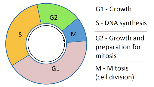
The cell cycle is an ordered set of events, culminating in cell growth and division into two daughter cells. It consists of four distinct phases: G1 (Gap 1), S (Synthesis), G2 (Gap 2), and M (Mitosis). These phases ensure that the cell grows, duplicates its DNA accurately, and divides this DNA equally between two daughter cells. Understanding these phases and their regulation is crucial for A-level biology, as it underpins not just how cells multiply, but also how they maintain genetic integrity and function in the organism.
Explanation of the Cell Cycle Phases: G1, S, G2, and M
- G1 Phase (Gap 1): This phase marks the beginning of the cell cycle. Cells increase in size, produce RNA, and synthesise proteins that are necessary for DNA replication. The G1 phase is crucial for the commitment to cell division, and its length can vary greatly among different cell types.
- S Phase (Synthesis): During the S phase, DNA replication occurs, ensuring that each daughter cell will receive an identical set of chromosomes. The cell duplicates its chromosomes, resulting in two sister chromatids joined at the centromere for each chromosome.
- G2 Phase (Gap 2): Following DNA synthesis, the cell enters the G2 phase, where further growth occurs. The cell synthesises proteins and continues to grow in preparation for mitosis. This phase also involves the critical process of DNA repair, ensuring any replication errors are corrected before cell division.
- M Phase (Mitosis): The M phase encompasses mitosis and cytokinesis. Mitosis is divided into several stages—prophase, metaphase, anaphase, and telophase—leading to the segregation of sister chromatids into two nuclei. Cytokinesis then divides the cell cytoplasm, resulting in two daughter cells.
Significance of Cell Cycle Regulation and Checkpoints
Regulation of the cell cycle is essential for the health and viability of organisms. The cell cycle is controlled by various checkpoints, which are mechanisms that ensure the cell is ready to proceed to the next phase. These checkpoints prevent errors such as DNA damage or incomplete DNA replication, which could lead to mutations or cell death.
- G1/S Checkpoint: Determines whether the cell has all the necessary resources and conditions to commit to DNA replication.
- G2/M Checkpoint: Assesses if DNA replication has been completed correctly without damage before the cell enters mitosis.
- Spindle Assembly Checkpoint (during Metaphase to Anaphase transition): Ensures that all chromosomes are properly attached to the spindle apparatus before chromatid separation.
These checkpoints are crucial for preventing the division of cells with damaged DNA or chromosomes that are not properly replicated, thereby maintaining genetic stability and preventing diseases like cancer. The cell cycle’s tight regulation ensures that cells divide only when conditions are optimal, highlighting the complexity and precision of cellular processes.

Kora F.
Maths | Chemistry | Biology Tutor
Student at UNIVERSITY OF LEEDS
£17 Per session
Book Free TrialUnderstanding the Cell Cycle
The cell cycle is a fundamental process through which cells ensure their growth, DNA replication, and division, facilitating the maintenance and development of organisms. This intricate cycle is divided into four main phases, each playing a vital role in preparing the cell for division and ensuring the faithful transmission of genetic information to daughter cells.
Explanation of the Cell Cycle Phases: G1, S, G2, and M
G1 Phase (Gap 1)
In the G1 phase, cells embark on a period of growth, accumulating the necessary resources for DNA replication and cell division. This phase involves significant biochemical activity, including the synthesis of proteins and RNA. The G1 phase is critical for the cell’s decision to commit to division, with various internal and external signals influencing its progression to the next phase, the S phase.
S Phase (Synthesis)
The S phase is characterized by the duplication of the cell’s DNA, ensuring that each daughter cell will inherit an identical set of chromosomes. This phase is marked by the replication of chromosomes, resulting in pairs of sister chromatids that are joined together at their centromeres. The accuracy of DNA replication during this phase is paramount for genetic fidelity.
G2 Phase (Gap 2)
Following DNA synthesis, the cell enters the G2 phase, a period of further growth and preparation for mitosis. The G2 phase allows the cell to repair any DNA damage that may have occurred during replication, ensuring the integrity of the genetic material. The synthesis of specific proteins required for mitosis also takes place during this phase.
M Phase (Mitosis)
The M phase encompasses the process of mitosis, where the replicated chromosomes are segregated into two new nuclei, and cytokinesis, which divides the cell’s cytoplasm, resulting in the formation of two separate daughter cells. Mitosis is further divided into stages: prophase, metaphase, anaphase, and telophase, each facilitating the orderly distribution of chromosomes.
Significance of Cell Cycle Regulation and Checkpoints
Cell cycle regulation involves a series of checkpoints that verify whether the cell is ready to progress to the next phase. These checkpoints play a crucial role in maintaining the cell’s genomic integrity, preventing the division of cells with damaged DNA or incomplete replication. The main checkpoints are:
- G1/S Checkpoint: Evaluates if the cell is ready for DNA replication.
- G2/M Checkpoint: Confirms the DNA is correctly replicated and free of damage before mitosis.
- Spindle Assembly Checkpoint: Ensures all chromosomes are correctly attached to the spindle before chromatid separation.
The precise regulation of the cell cycle and its checkpoints underscores the complexity of cellular processes, safeguarding the organism’s health by preventing the proliferation of damaged cells, thereby averting potential diseases, including cancer.
Detailed Phases of Mitosis
Mitosis is a critical process in the cell cycle, allowing for the division of a single cell into two genetically identical daughter cells. This section delves into the stages of mitosis, highlighting their significance and the intricate events that occur at each phase to ensure accurate and efficient cell division.
Introduction to the Stages of Mitosis and Their Significance
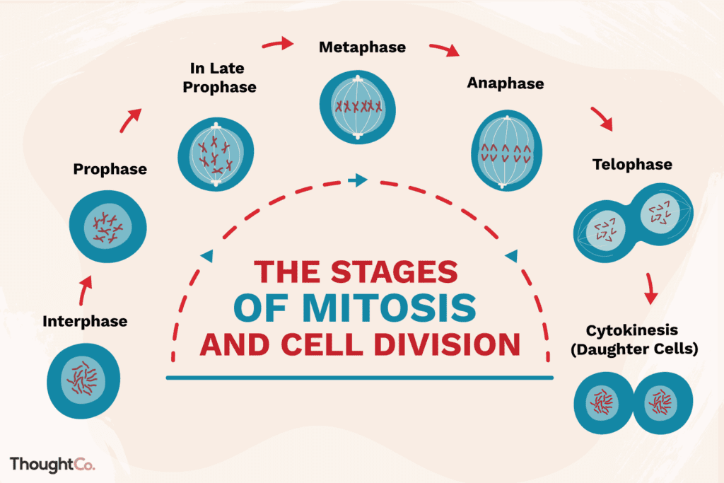
Mitosis is divided into distinct stages: prophase, prometaphase, metaphase, anaphase, and telophase, each with specific functions and processes that ensure the DNA is correctly replicated and distributed. Understanding these stages is crucial for comprehending how cells divide and the mechanisms that maintain genetic consistency across cell generations.
Interphase
While not a part of mitosis itself, interphase is the phase preceding mitosis, where the cell prepares for division. This stage is characterized by critical activities that set the stage for successful cell division.
G1 Phase (Gap 1)
Cells grow and synthesise proteins necessary for DNA replication, laying the groundwork for the S phase.
S Phase (Synthesis)
DNA replication occurs, ensuring each chromosome is duplicated to produce sister chromatids, vital for genetic equality in daughter cells.
G2 Phase (Gap 2)
Further cell growth and protein synthesis occur, with crucial DNA repair mechanisms ensuring the integrity of the replicated DNA.
The importance of DNA replication during interphase cannot be overstated, as it ensures each daughter cell receives an exact copy of the genetic material.
Prophase
Prophase marks the beginning of mitosis, characterized by several key events:
- Chromatin Condensation: Chromatin fibers condense into visible chromosomes, each consisting of two identical sister chromatids joined at the centromere.
- Centriole Movement: In animal cells, centrioles begin to move to opposite ends of the cell, initiating the formation of the mitotic spindle.
- Spindle Fibre Formation: The mitotic spindle, a structure made of microtubules, begins to form between the moving centrioles.
- Nuclear Envelope Breakdown: The nuclear envelope disassembles, allowing the spindle fibers to contact the chromosomes.
These changes prepare the cell for the segregation of chromosomes to opposite poles.
Prometaphase
Prometaphase follows prophase and involves further development of the structures necessary for chromosome movement:
- Spindle Fibres Attach to Kinetochores: Spindle fibers attach to protein structures called kinetochores located at the centromeres of each chromosome.
- Chromosomes Begin Moving: The attachment of spindle fibers to kinetochores facilitates the movement of chromosomes toward the cell’s equator, setting the stage for their alignment during metaphase.
The introduction of prometaphase into the discussion provides a more detailed understanding of the transition between the initial chromosome condensation and their eventual alignment along the metaphase plate.
Metaphase
Metaphase is a critical stage in mitosis where chromosomes align at the metaphase plate, an imaginary line equidistant between the spindle’s two poles. This alignment ensures that each daughter cell will receive an identical set of chromosomes.
- Chromosomes Align at the Metaphase Plate: Each chromosome, attached to spindle fibres via its kinetochore, is aligned precisely at the cell’s equator. This precise alignment is crucial for the equal distribution of genetic material.
- The Role of Spindle Fibres in Alignment: Spindle fibres play a vital role in chromosome alignment by exerting forces that pull the chromosomes to the centre of the cell. This balance of forces ensures that each sister chromatid is equally positioned between the two poles of the cell.
Anaphase
Anaphase begins when the sister chromatids, previously held together at the centromere, separate and are pulled towards opposite poles of the cell. This phase is characterized by two key events:
- Sister Chromatids Separate and Move to Opposite Poles: Once all chromosomes are correctly aligned, the centromeres split, allowing the sister chromatids to be pulled apart. Motor proteins along the spindle fibres facilitate this movement, ensuring that each pole receives an identical set of chromosomes.
- Key Molecular Mechanisms Driving Chromatid Separation: The separation of sister chromatids is driven by the shortening of spindle fibres and the action of motor proteins. Additionally, enzymes that degrade cohesin, the protein holding sister chromatids together, are activated, allowing the chromatids to separate.
Telophase
Telophase is the final stage of mitosis, marked by the re-establishment of normal cell structures and the beginning of cytokinesis (the division of the cell’s cytoplasm).
- Chromatids Arrive at Poles and Begin to Decondense: Upon reaching the poles, the chromatids, now individual chromosomes, begin to decondense, returning to a less compact chromatin state. This decondensation allows for the re-initiation of normal nuclear activities.
- Reformation of the Nuclear Envelope: Around each set of chromosomes, a new nuclear envelope forms, re-establishing the nuclei. This reformation is crucial for segregating the genetic material into two distinct nuclei.
- Characteristics of the Newly Formed Nuclei: The newly formed nuclei contain a complete set of chromosomes identical to those of the parent cell. Nucleoli reappear, and the chromosomes gradually decondense, signaling the end of mitosis and the transition into the interphase of the next cell cycle.
These stages of mitosis ensure that the cell’s genetic material is accurately and equally divided, allowing for the production of two genetically identical daughter cells. Understanding these processes is essential for grasping how cells replicate and maintain genetic continuity through generations.
Cytokinesis: Division of the Cytoplasm

Cytokinesis is the process that follows the completion of mitosis, marking the physical separation of the cytoplasm into two distinct daughter cells. This phase is crucial for ensuring that the newly formed nuclei are housed in separate cells, each with its own complete set of cellular machinery and membrane. The mechanism of cytokinesis differs significantly between animal and plant cells, reflecting the unique structural challenges presented by their respective cell types.
Mechanism of Cytoplasmic Division in Animal and Plant Cells
In animal cells, cytokinesis is accomplished through a process known as cleavage. This involves the formation of a cleavage furrow, an indentation of the cell membrane around the equator of the cell. The furrow deepens as a ring of actin and myosin filaments contracts, pulling the plasma membrane inward. This contraction continues until the membrane is pinched off, separating the cell into two daughter cells, each with its own nucleus and cytoplasm.
In plant cells, the presence of a rigid cell wall prevents the formation of a cleavage furrow. Instead, cytokinesis is achieved through the construction of a cell plate in the middle of the cell. Vesicles from the Golgi apparatus, filled with cell wall materials, coalesce at the site of division inside the cell. These vesicles fuse to form the cell plate, which gradually expands outward until it merges with the cell wall, effectively dividing the cell into two. Each daughter cell then builds its own new cell wall segment along the sides of the cell plate.
Differences Between Plant and Animal Cytokinesis
The key differences between cytokinesis in plant and animal cells are driven by their structural differences, notably the presence of a cell wall in plant cells:
- Mechanism of Division: Animal cells use a contractile ring to pinch the cell in two, while plant cells build a new dividing wall to separate the daughter cells.
- Structural Challenges: The rigid cell wall in plant cells necessitates the construction of a cell plate from the inside out, as opposed to the contractile mechanism in animal cells, which can directly constrict the cell membrane.
- Components Involved: In animal cells, actin and myosin filaments drive the constriction of the cleavage furrow. In contrast, plant cells rely on vesicles from the Golgi apparatus to supply the materials needed for cell plate formation.
Understanding these processes is essential for a comprehensive grasp of cell division and the physical separation of cells following mitosis. Cytokinesis ensures that each daughter cell receives not only a complete set of genetic information but also a sufficient amount of cytoplasm and organelles, ready to function as independent entities.

Regulation of Mitosis
The process of mitosis is tightly controlled by various checkpoints and regulatory proteins to ensure that cell division proceeds correctly and at the appropriate time. This regulation is crucial for maintaining the health and stability of an organism, as it prevents the unchecked cell growth that can lead to cancer and other diseases.
Importance of Checkpoints and Regulatory Proteins
Mitosis is regulated by specific checkpoints within the cell cycle that assess whether the cell is ready to proceed to the next phase. These checkpoints serve as critical control points where the cell evaluates the integrity of its DNA, the success of replication, and the proper alignment of chromosomes. Regulatory proteins, including cyclins and cyclin-dependent kinases (CDKs), play a pivotal role in this process. They act as signals that prompt the cell to proceed with mitosis, or halt the cycle to allow for repair or growth.
- G1/S Checkpoint: Ensures that the cell’s DNA is undamaged and the cell is sufficiently grown before DNA replication begins.
- G2/M Checkpoint: Verifies that DNA has been completely and accurately replicated and that the cell is ready to enter mitosis.
- Spindle Assembly Checkpoint (SAC): Checks that all chromosomes are correctly attached to the spindle apparatus before allowing the cell to proceed from metaphase to anaphase, ensuring accurate chromosome segregation.
These checkpoints prevent the division of cells that have damaged DNA or other cellular defects, thereby safeguarding the organism’s genetic integrity.
Consequences of Dysregulation: Cancer and Other Diseases
Dysregulation of the cell cycle can lead to uncontrolled cell division, contributing to the development of cancer and other diseases. Mutations in the genes that encode regulatory proteins can lead to the loss of checkpoint control, allowing cells with damaged DNA or incomplete replication to divide. This can result in the accumulation of genetic mutations and the formation of tumors.
- Cancer: Cancer arises from the uncontrolled division of abnormal cells. Mutations in regulatory genes can cause cells to bypass checkpoints, leading to the proliferation of cells with genetic errors.
- Other Diseases: Besides cancer, dysregulation of mitosis can contribute to a range of diseases, including developmental disorders and conditions characterized by abnormal cell growth or death.
Understanding the regulation of mitosis and the mechanisms that control cell division is essential for developing treatments for cancer and other diseases resulting from cell cycle dysregulation. Targeting specific molecules involved in cell cycle regulation offers a promising approach for therapeutic intervention, aiming to restore normal cell division and prevent the growth of tumors.
Mitotic Index and Its Applications
The mitotic index is a quantitative measure that reflects the percentage of cells in a given population undergoing mitosis at a specific time. This index is a crucial parameter in both research and clinical settings, as it provides insight into the proliferation rate of a cell population, which can be indicative of the tissue’s growth or health status.
How to Calculate and Interpret the Mitotic Index
The mitotic index is calculated by dividing the number of cells in mitosis by the total number of cells observed, then multiplying by 100 to obtain a percentage:
Mitotic Index=(Total number of cells observed/Number of cells in mitosis)×100
To accurately calculate this index, a representative sample of cells is stained and examined under a microscope. Cells in different stages of mitosis (prophase, metaphase, anaphase, telophase) are counted, along with the total number of cells, to determine the proportion of cells that are actively dividing.
Interpreting the mitotic index involves understanding its implications for cell growth and proliferation. A high mitotic index indicates a high rate of cell division, which is typical in rapidly growing tissues, such as embryonic tissues or tumours. Conversely, a low mitotic index suggests slower cell division, common in mature or differentiated tissues.
The Relevance of Mitotic Index in Medical Diagnosis and Research
The mitotic index is particularly valuable in oncology, where it is used to assess the aggressiveness of tumours. Tumours with a high mitotic index are often more aggressive and have a poorer prognosis than those with a lower index, as rapid cell division suggests a high potential for growth and spread. This information can be critical for determining the most appropriate treatment strategy, including the need for aggressive therapy.
In research, the mitotic index is used to study cell cycle dynamics and the effects of various substances on cell proliferation. It can help in identifying potential anticancer drugs by evaluating their ability to inhibit cell division. Moreover, in tissue engineering and stem cell research, the mitotic index is used to monitor the growth rate of cells, ensuring that they are proliferating at a desired rate for therapeutic applications.
Overall, the mitotic index serves as a vital tool in both the diagnosis and study of diseases, particularly cancer, offering insights into cell proliferation that are crucial for effective treatment and understanding of cellular dynamics.
Conclusion
Understanding the mechanisms of mitosis and the cell cycle is not just about memorizing stages or recognising cellular structures under a microscope. It’s about appreciating the complex dance of life at its most fundamental level. These processes are the foundation of growth, development, and the maintenance of life itself. From the precise orchestration of chromosomes during mitosis to the critical regulation of the cell cycle, every aspect reflects the remarkable efficiency and adaptability of cellular machinery.
The study of mitosis offers more than just insights into cellular division; it opens a window into the processes that drive life, the errors that can lead to disease, and the potential for therapeutic intervention. It challenges us to think about how life proliferates, repairs, and evolves at the microscopic level. Moreover, it underscores the importance of rigorous scientific inquiry and the continuous search for understanding that drives biology forward.
In the context of A-level biology, grasping these concepts is not just about academic achievement; it’s about building a foundation for future scientific exploration and understanding the biological principles that underpin health and disease. Whether aiming to pass or secure an A*, students can find themselves navigating complex topics that demand both memorization and deep understanding.
For those seeking to enhance their learning journey, support is available. Edumentors offers access to top tutors from leading UK universities, individuals who have not only excelled in their exams but also possess a passion for sharing their knowledge and strategies for success. These A-level biology tutors, having achieved the highest grades themselves, are uniquely positioned to provide guidance, insights, and the tailored support students need to excel in A-level biology and beyond. Engaging with Edumentors could be the step that transforms ambition into achievement, opening doors to further education and careers in the vast world of biology.


